Identifying bones and bone markings on radiographs of skull – Delving into the intricate realm of identifying bones and bone markings on radiographs of the skull, this comprehensive guide unravels the complexities of this essential skill. Through a meticulous examination of the skull’s anatomy and the interpretation of radiographic images, this guide empowers readers with the knowledge and expertise to navigate the intricacies of this field.
By providing a comprehensive overview of the different bones that constitute the skull, their location, shape, and function, this guide lays the foundation for understanding the structural framework of the skull. Moreover, it delves into the diverse types of bone markings found on the skull, explaining their significance and providing examples of their location.
Identifying Bones of the Skull

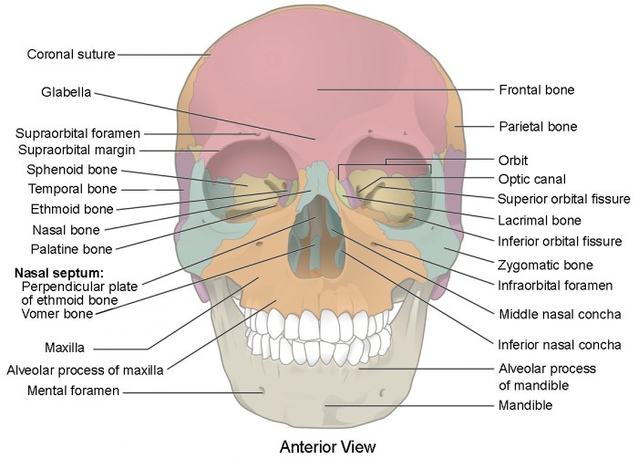
The skull is composed of 22 bones, which can be divided into two groups: the cranial bones and the facial bones. The cranial bones form the protective casing for the brain, while the facial bones form the framework of the face.
Cranial Bones, Identifying bones and bone markings on radiographs of skull
The cranial bones are eight in number and include the frontal bone, parietal bones, occipital bone, temporal bones, sphenoid bone, and ethmoid bone. The frontal bone forms the forehead and the roof of the orbits. The parietal bones form the sides and roof of the skull.
The occipital bone forms the back of the skull and the base of the cranium. The temporal bones form the sides of the skull and contain the organs of hearing and balance. The sphenoid bone forms the base of the skull and contains the pituitary gland.
The ethmoid bone forms the roof of the nasal cavity and contains the olfactory bulbs.
Facial Bones
The facial bones are 14 in number and include the nasal bones, maxillae, zygomatic bones, lacrimal bones, palatine bones, inferior nasal conchae, vomer, and mandible. The nasal bones form the bridge of the nose. The maxillae form the upper jaw and contain the teeth.
The zygomatic bones form the cheekbones. The lacrimal bones form the medial walls of the orbits. The palatine bones form the posterior part of the hard palate. The inferior nasal conchae are scroll-like bones that project into the nasal cavity.
The vomer is a thin bone that forms the nasal septum. The mandible is the lower jaw and contains the teeth.
| Bone Name | Location | Shape | Function |
|---|---|---|---|
| Frontal bone | Forehead and roof of orbits | Flat and square | Protects the brain |
| Parietal bones | Sides and roof of skull | Convex and quadrilateral | Protects the brain |
| Occipital bone | Back of skull and base of cranium | Flat and triangular | Protects the brain and supports the head |
| Temporal bones | Sides of skull | Irregular and complex | Contains organs of hearing and balance |
| Sphenoid bone | Base of skull | Butterfly-shaped | Forms pituitary gland and supports other cranial bones |
| Ethmoid bone | Roof of nasal cavity | Thin and scroll-like | Forms olfactory bulbs and supports other facial bones |
| Nasal bones | Bridge of nose | Flat and rectangular | Protects the nasal cavity |
| Maxillae | Upper jaw | Large and complex | Contains teeth and forms hard palate |
| Zygomatic bones | Cheekbones | Triangular and flat | Forms cheekbones and supports maxillae |
| Lacrimal bones | Medial walls of orbits | Small and thin | Forms medial walls of orbits and supports lacrimal glands |
| Palatine bones | Posterior part of hard palate | Thin and L-shaped | Forms posterior part of hard palate and supports maxillae |
| Inferior nasal conchae | Nasal cavity | Scroll-like and thin | Directs airflow through nasal cavity |
| Vomer | Nasal septum | Thin and triangular | Forms nasal septum and supports other facial bones |
| Mandible | Lower jaw | U-shaped and robust | Contains teeth and supports lower face |
Identifying Bone Markings on the Skull

Bone markings are anatomical features that can be found on the surface of bones. They serve a variety of functions, such as providing attachment points for muscles, ligaments, and tendons, and allowing for the passage of nerves and blood vessels.
There are many different types of bone markings, and each type has a specific function.
Types of Bone Markings
- Sutures: Immovable joints between the bones of the skull. They are filled with fibrous tissue and allow for some movement of the bones during growth.
- Foramina: Holes or openings in the skull that allow for the passage of nerves and blood vessels.
- Fissures: Grooves in the skull that allow for the passage of nerves and blood vessels.
- Processes: Projections from the surface of the skull that provide attachment points for muscles, ligaments, and tendons.
- Sinuses: Air-filled cavities within the skull that help to reduce the weight of the skull and provide resonance for the voice.
Functions of Bone Markings
- Provide attachment points for muscles, ligaments, and tendons
- Allow for the passage of nerves and blood vessels
- Reduce the weight of the skull
- Provide resonance for the voice
Using Radiographs to Identify Bones and Bone Markings

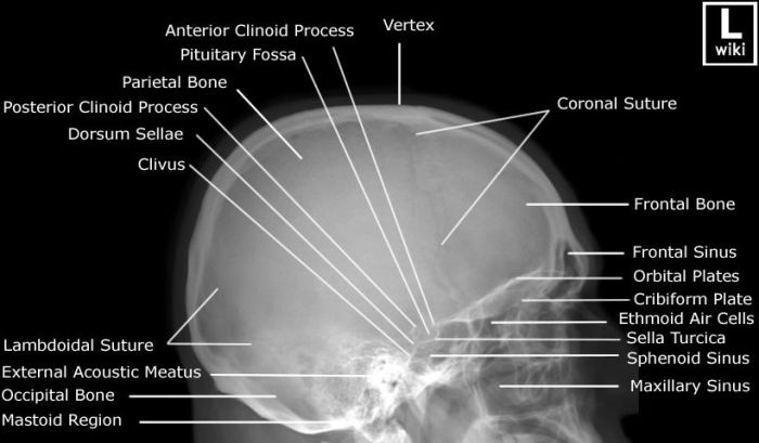
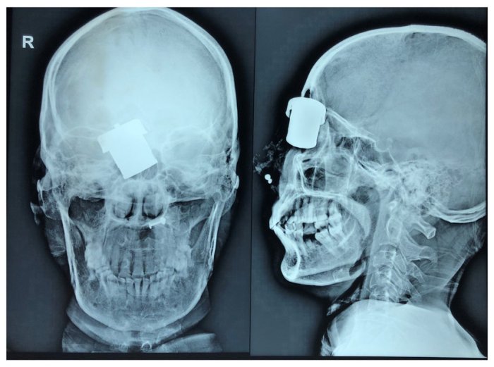
Radiographs are X-ray images that can be used to visualize the bones and bone markings of the skull. Radiographs are commonly used to diagnose fractures, tumors, and other abnormalities of the skull.
Types of Radiographs
- Plain radiographs: These are the most common type of radiograph and provide a general view of the skull.
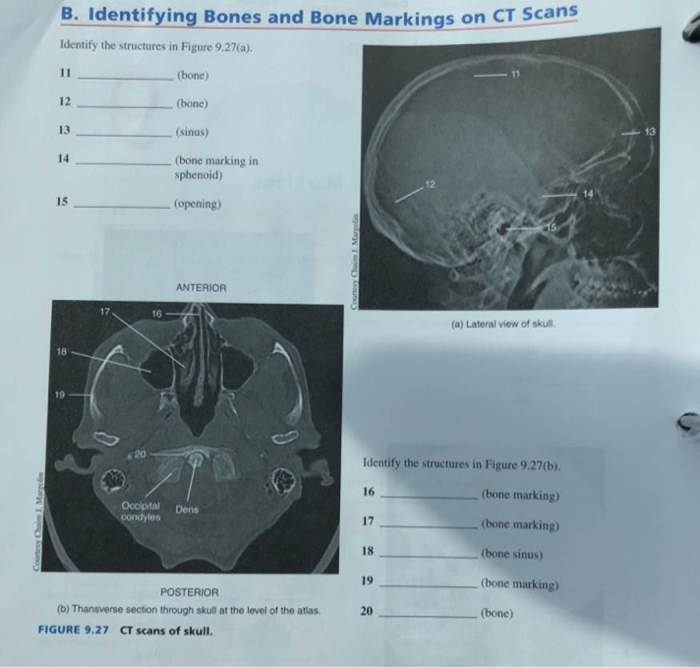
- Computed tomography (CT) scans: These are more detailed than plain radiographs and can provide cross-sectional images of the skull.
- Magnetic resonance imaging (MRI) scans: These are the most detailed type of radiograph and can provide images of the soft tissues of the skull as well as the bones.
Identifying Bones and Bone Markings on Radiographs
When identifying bones and bone markings on radiographs, it is important to consider the following factors:
- The location of the bone or bone marking
- The shape of the bone or bone marking
- The density of the bone or bone marking
- The presence of any abnormalities
By considering these factors, it is possible to accurately identify bones and bone markings on radiographs.
Clinical Applications of Identifying Bones and Bone Markings on Radiographs of the Skull

Identifying bones and bone markings on radiographs of the skull is a valuable tool for diagnosing and treating a variety of conditions. Some of the most common clinical applications include:
- Diagnosing fractures: Radiographs can be used to identify fractures of the skull, which can be caused by trauma or disease.
- Diagnosing tumors: Radiographs can be used to identify tumors of the skull, which can be benign or malignant.
- Diagnosing infections: Radiographs can be used to identify infections of the skull, such as osteomyelitis.
- Planning surgery: Radiographs can be used to plan surgery on the skull, such as surgery to repair a fracture or remove a tumor.
- Monitoring treatment: Radiographs can be used to monitor the progress of treatment for a variety of conditions, such as fractures, tumors, and infections.
Questions Often Asked: Identifying Bones And Bone Markings On Radiographs Of Skull
What are the different types of bone markings found on the skull?
Bone markings include sutures, foramina, processes, and fossae, each serving specific functions in the skull’s anatomy.
How are radiographs used to identify bones and bone markings?
Radiographs provide a non-invasive method to visualize the skull’s internal structures, allowing for the identification of bones and bone markings based on their radiographic appearance.
What are the clinical applications of identifying bones and bone markings on radiographs of the skull?
Identifying bones and bone markings on radiographs aids in diagnosing and treating various conditions, such as fractures, tumors, and congenital anomalies, guiding appropriate medical interventions.